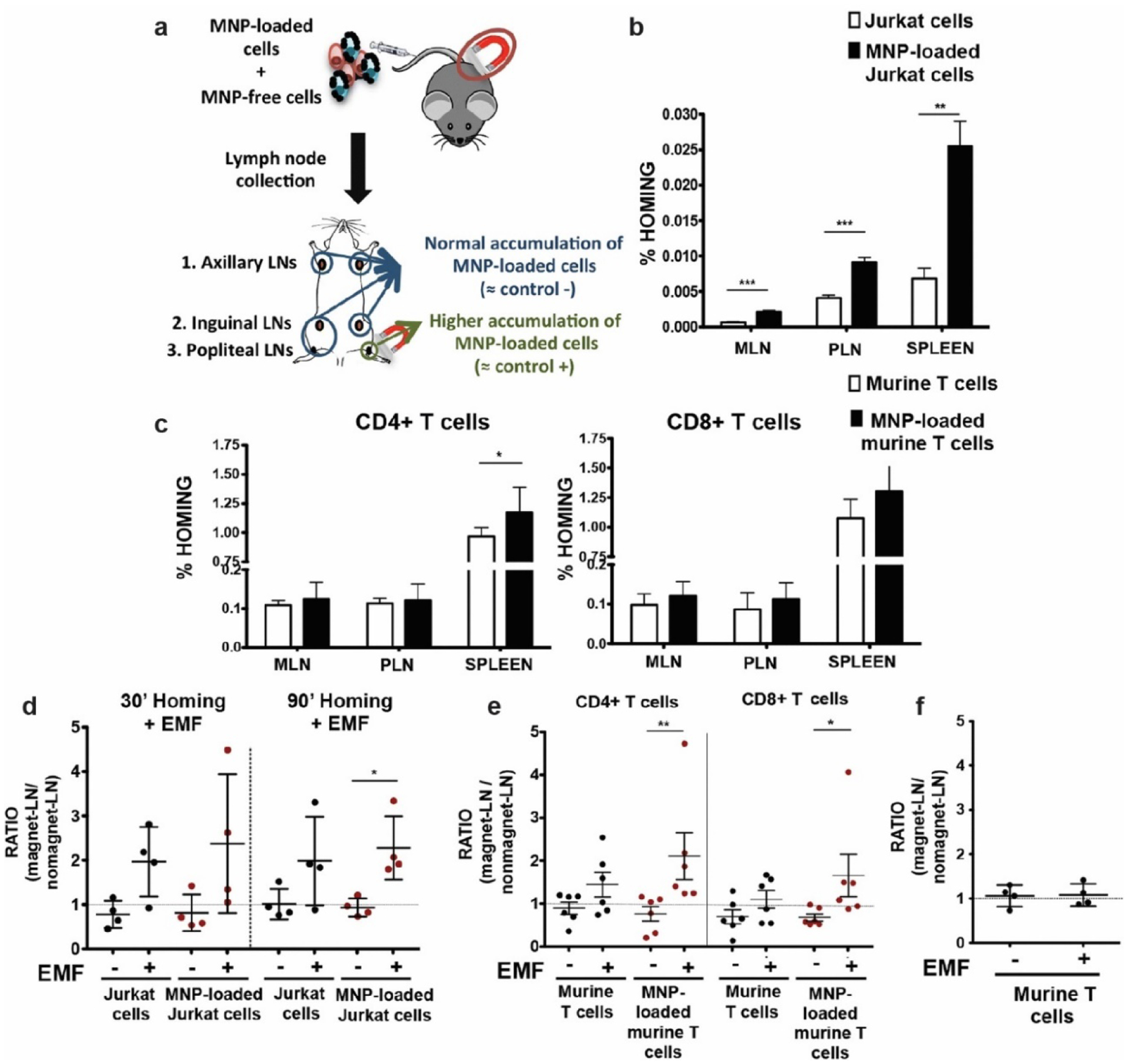
Figure 8.

In vivo homing capacity of Jurkat human T cells and murine primary T cells after magnetic iron oxide nanoparticle (MNP) treatment with and without an external magnetic field (EMF). (a) Experimental set-up for determining the homing capacity of MNP-loaded cells compared to MNP-free cells. A mixture of differentially fluorescence-labelled MNP-free and MNP-loaded Jurkat or murine T cells was prepared and intravenously injected into nude (Jurkat) or C57BL/6J (murine T cells) recipient mice. After 1 h, peripheral (PLN) and mesenteric (MLN) lymph nodes (LN) and spleen were collected, processed, and analyzed by flow cytometry. Homing capacity of MNP-free and MNP-loaded (b) Jurkat and (c) murine T cells in the absence of an EMF, 1 h after cell injection. Ratio of MNP-free and MNP-loaded (d) Jurkat and (e) murine T cells in the LNs exposed to an EMF to control LN (no EMF), 20 min after intravenous injection of the cell mixture into recipient mice, normalized to the input ratio. (f) Ratio of MNP-free murine T cells, administered alone as control, in the LN exposed to an EMF to control LN (no EMF) after intravenous injection. Reproduced with permission.126 Copyright 2019, Springer Nature.
