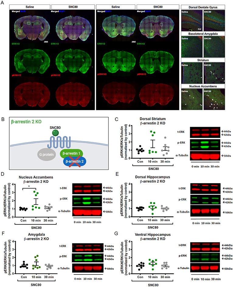
Figure 3. ERK1/2 activation in the amygdala and the dorsal hippocampus are β-arrestin 2 dependent.
(A) for ERK (green), phosphorylated ERK (red), and nuclear DNA marker DAPI (blue) in limbic and striatal regions of β-arrestin 2 KO mouse brain tissues that were perfused 10 min after i.p. administration with saline or SNC80 (20 mg/kg). 10x-Magnification images (left; scale bars, 1000 μm) were used to stitch together the whole brain-slice images shown. Enlarged images (right; scale bars, 100 μm) are 20x magnification that correspond to the regions marked (white boxes) in the 10x images. (B) Schematic of the cellular context in β-arrestin 2 KO mice. (C to G) Blotting analysis of SNC80-induced ERK1/2 activation in the dorsal striatum (C), nucleus accumbens (D), dorsal hippocampus (E), amygdala (F), and ventral hippocampus (G) at 10 and 30 minutes after i.p. administration with SNC80 (20 mg/kg). Con, 0 min: saline administered. Data are from N = 7 to 8 mice; see table S2. Representative blots are shown to the right of the related bar graph. See table S2 for one-way ANOVA and post-hoc multiple comparisons.

