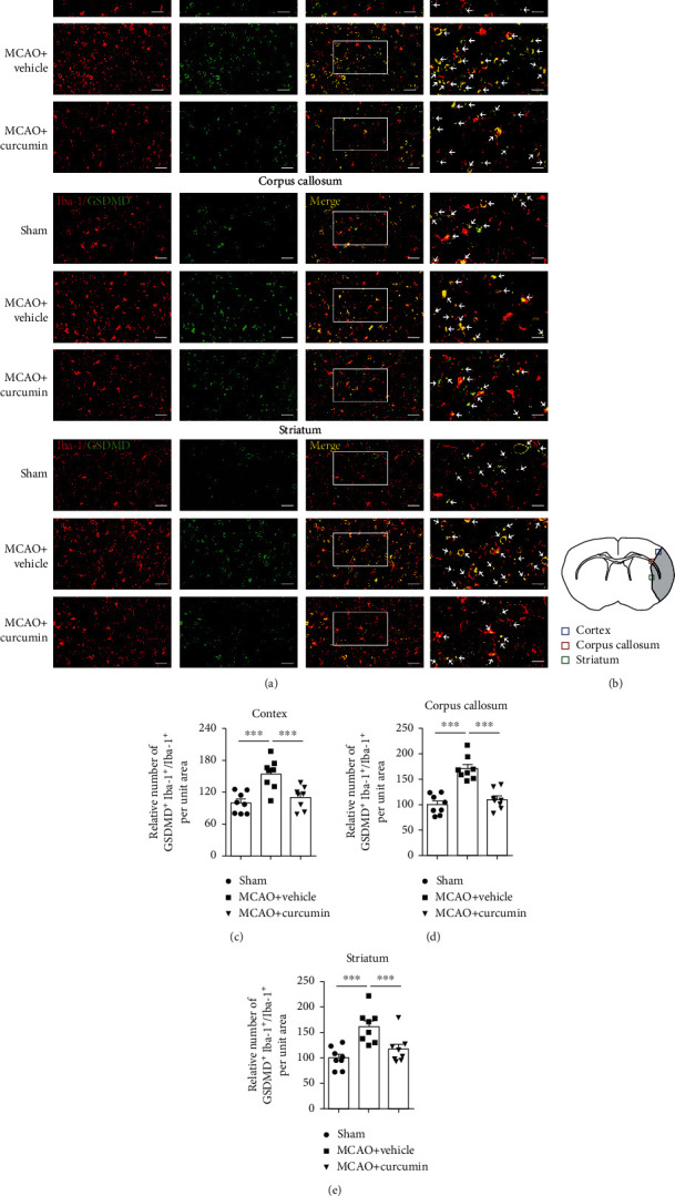
Figure 3.

Curcumin treatment reduces GSDMD-positive microglia in the ischemic cortex, corpus callosum, and striatum 21 days after MCAO. (a) Representative coimmunostaining of Iba-1 (red) and GSDMD (green). Scale bar in the figures of 1-3 columns from left = 20 μm, and scale bar in the figure of the 4th column from left = 10 μm. Arrows indicate GSDMD+Iba-1+ microglia. (b) Schematic diagram illustrating the anatomical location of images in the ipsilateral perilesion cortex (blue), corpus callosum (red), and striatum (green). (c–e) Quantitative analysis of GSDMD-positive microglia. Quantification of the percentage of GSDMD+Iba-1+ cells among total Iba-1+ cells in the perilesion cortex (c), corpus callosum (d), and striatum (e). Values are the mean ± SEM. ∗∗∗p < 0.001. n = 8 mice per group. One-way ANOVA followed by Bonferroni post hoc test.
