Figure 6.

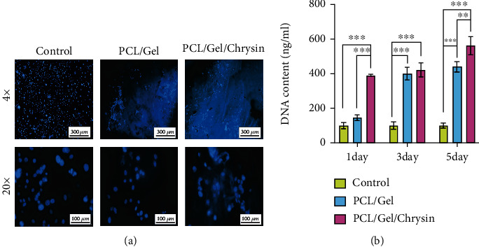
(a) The cell viability of the DPSCs by DAPI staining in control, PCL/Gel, and PCL/Gel/Chyrsin groups for 5 days. (b) The DNA content extracted from DPSCs grown into control, PCL/Gel, and PCL/Gel/Chrysin on days 1, 3, and 5.

(a) The cell viability of the DPSCs by DAPI staining in control, PCL/Gel, and PCL/Gel/Chyrsin groups for 5 days. (b) The DNA content extracted from DPSCs grown into control, PCL/Gel, and PCL/Gel/Chrysin on days 1, 3, and 5.