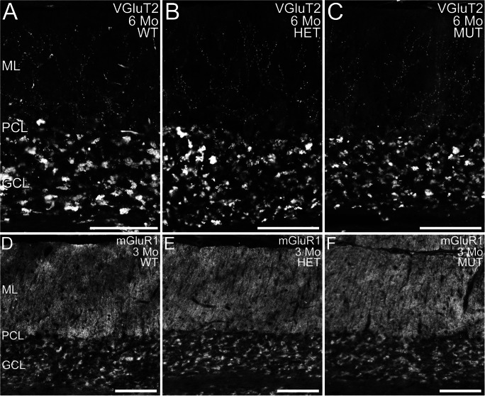
Fig. 5.
Distribution of VGluT2 and mGluR1 in WT, HET, and MUT rat cerebellum. (A–C) Distribution of VGluT2, a marker for climbing fiber synapses, is comparable in the cerebellar cortex of WT, HET, and MUT rats. Labeling is present in climbing fiber synapses onto Purkinje cell dendrites in the molecular layer (ML) and in the synaptic glomeruli of the granule cell layer (GCL). PCL, Purkinje cell layer. (D–F) Distribution of mGluR1 is comparable in the WT, HET, and MUT cerebellum. PCL, Purkinje cell layer. Scale bars = 100 μm for panels (A–C); 200 μm for panels (D–F)

