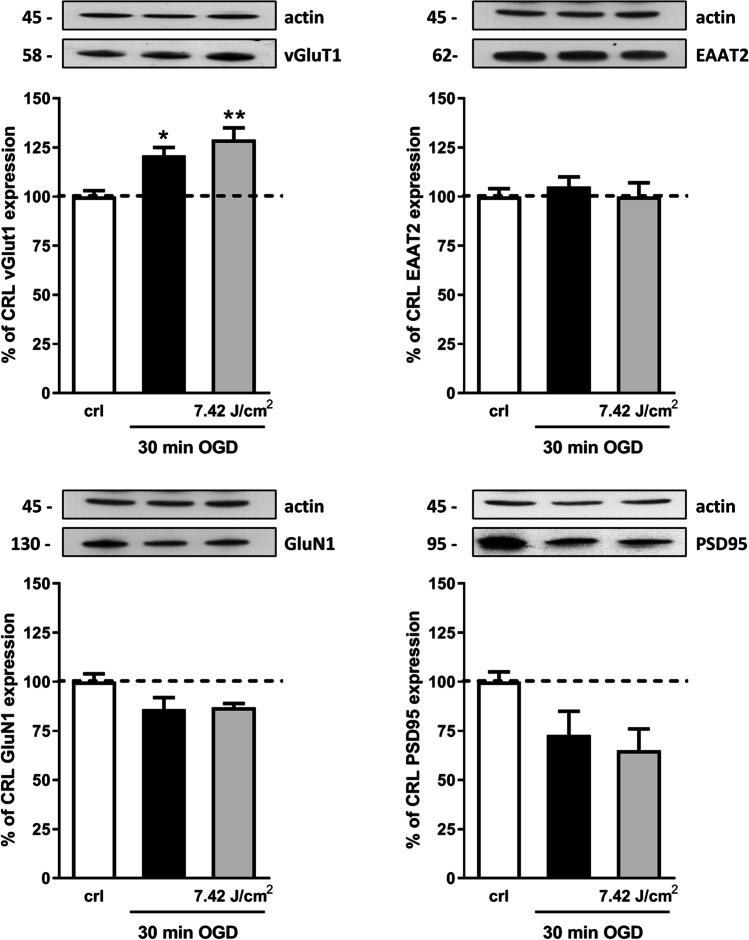
Fig. 4.
Effects of NIR laser treatment after OGD toxicity on proteins involved in excitatory synaptic activity. Experiments were conducted as described in Fig. 1. Hippocampal slices were exposed to 30 min OGD and immediately after to 7.42 J/cm2 NIR laser. Twenty-four hours later, the expression of GluN1, PSD95, vGluT1, and EAAT2 was evaluated in total homogenate by Western blot analysis. Data are expressed as percentage of control. Bars represent the mean ± SEM of at least three experiments run in ottuplicate. *P < 0.05, **P < 0.01 vs. CRL, #P < 0.05, ##P < 0.01 vs. OGD (ANOVA + Tukey’s w test)

