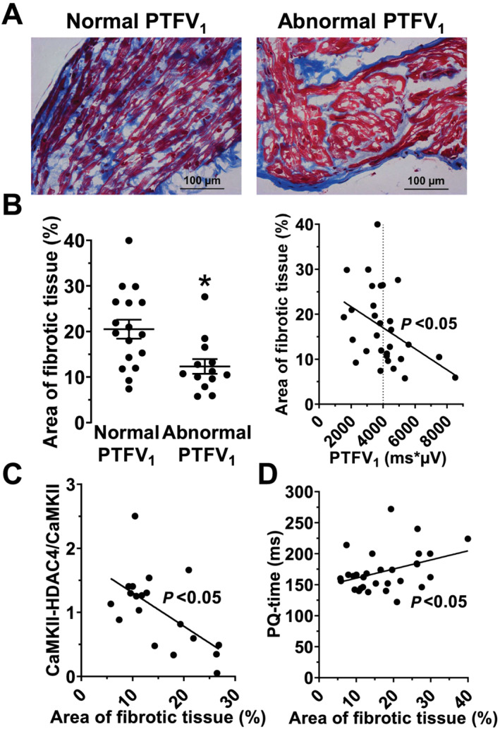
Figure 3.

Atrial fibrosis is decreased in patients with an abnormal PTFV1. (A) Original micrographs showing myocardial fibrosis (Masson's trichrome staining) in trabeculae of CABG patients with a normal and an abnormal PTFV1 (400× magnification). (B) Interestingly, the area of myocardial fibrosis (%) was significantly reduced in patients with an abnormal PTFV1 (n = 13 vs. 17), leading to a significant negative correlation with the PTFV1 (n = 30). (C) Moreover, we also found the area of fibrosis correlating significantly negative with the CaMKII activity, indicating that active CaMKII can only be found in cardiomyocytes and not in fibrotic tissue (n = 19). (D) Additionally, we found the area of fibrosis correlating significantly positive with a slowed atrial conduction velocity (i.e. PQ time, n = 30). *P < 0.05, Mann–Whitney test and linear regression analysis, as appropriate.
