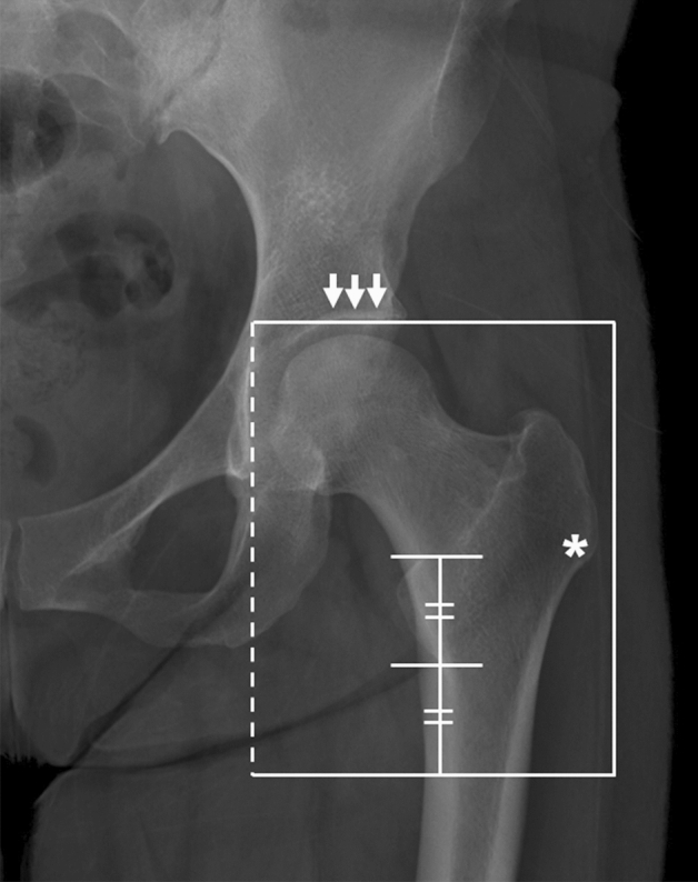
Figure 4.

The region of interest that was cropped on simple hip radiograph. The upper border of the cropped image was the acetabular roof (arrows) and femoral head, and the lower border was located as far as the length of the lesser trochanter from the tip of it. The medial border (dotted line) was the crossing of the lateral margin of teardrop, and the lateral border of cropped image was positioned just lateral of the vastus ridge (asterisk).
