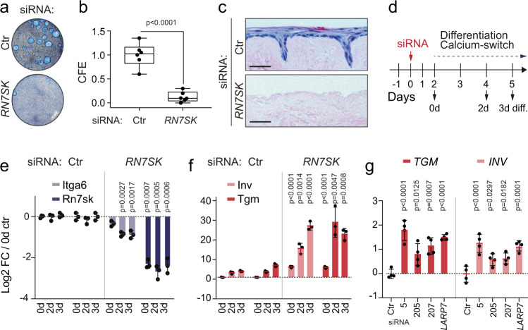
Fig. 2. RN7SK-depletion induces differentiation of primary human keratinocytes.
a, b Representative image (a) and quantification (b) of the colony-forming assay (CFE) using primary human keratinocytes transfected with control (Ctr) or RN7SK siRNAs (n = 6 transfections). Box plots show all data points, median, interquartile range, and whiskers ending at minimum and maximum values. c H&E staining of human skin reconstituted ex vivo on de-epidermalised dermis (DED) using primary human keratinocytes transfected with Ctr or RN7SK siRNAs. Shown is one representative image out of two DED assays. Scale bars: 10 μm. d–g Treatment regime in days (d) and log2 fold-change (FC) of RNA levels of Integrin α6 (ITGA6; light blue) and RN7SK (dark blue) (e), or Transglutaminase 1 (TGM; red) and Involucrin (INV; pink) (f) in transfected human keratinocytes. (n = 3 transfections). g Log2 FC of Tgm and Inv RNA levels in human keratinocytes 48 h after transfection of the indicated siRNAs (n = 4 transfections). 5, 205, and 207: different siRNAs targeting RN7SK. Data are normalized to GAPDH (e–g). Shown is mean ± SD (e−g). Two-tailed unpaired t-test (b). One-way ANOVA (e−g). Exact p-values are indicated. Source data are provided as a Source Data file.

