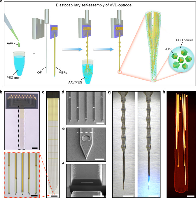
Fig. 1. Self-assembled multifunctional VVD-optrodes.
a Schematics of the elastocapillary self-assembly process of a VVD-optrode. b A MEF array. Scale bar, 2 mm. c Zoom-in views of the plane-mesh-filamentous structure in the black dashed box in (b). Scale bar, 1 mm and 50 μm (magnified view). d SEM image (tilted at 45°) of the microelectrode filaments of a MEF array. Scale bar, 50 μm. e Enlarged SEM image (tilted at 45°) of a microelectrode filament. Scale bar, 10 μm. f Focused ion beam-polished cross-section along the white dashed line in (e). Scale bar, 5 μm. g A self-assembled VVD-optrode without (left) and with blue light illumination (right), respectively. Scale bar, 1 mm. h False-color micro-CT image of a self-assembled probe. Scale bar, 50 μm.

