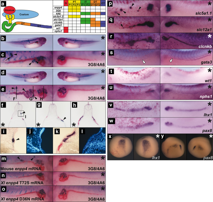
Fig. 1. Overexpression of enpp4 induces ectopic proximal pronephric tubules.
a Schematic diagram of pronephric structural components showing the expression domain for each marker used in this study, adapted from ref. 21. G: glomus, PT: proximal tubule, IT: intermediate tubule, DT: distal tubule, CT: collecting tubule. b–y Embryos injected with 2 ng of enpp4 and 250 pg of LacZ mRNAs were examined by 3G8/4A6 antibody staining (b–o) or whole-mount in situ hybridization with the following probes: slc5a1.1 (p), slc12a1 (q), clcnkb (r) and gata3 (s) at stage 37/38; wt1 (t) and nphs1 (u) at stage 32; lhx1 (v, x) and pax8 (w, y) at stages 28 and 14. f–l Transverse sections of the embryo shown in panels (d) and (e) were cut in the anterior–posterior registers indicated by lines in panel (e). A higher magnification image (i) of ectopic pronephros in the somite indicated by square in (f) and of control kidney (k) and counterstained with Hoechst to indicate nuclei (j, l). Embryos injected with 2 ng of mouse wild-type Enpp4 (m), X. laevis mutated in the putative catalytic site (n) or in the cation binding site (o) and 250 pg of LacZ mRNAs were examined by 3G8/4A6 antibody staining. The asterisk denotes the uninjected side of each embryo. Arrowheads indicate ectopic marker staining. Blank arrowheads in (s) indicate the anterior limit of gata3 expression. See also Supplementary Table 1 for raw data and statistical analyses and Supplementary Fig. 1.

