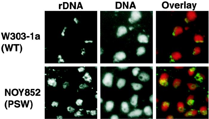
FIG. 7.
FISH analysis of rDNA in PSW strain NOY852 and control strain W303-1a. Yeast strains were analyzed for rDNA and DNA as described in Materials and Methods. Images of rDNA and DNA were pseudocolored green and red, respectively, giving overlapped regions yellow in overlay. Individual images are shown in black and white. Note that in the PSW strain, many of the DAPI-stained nuclei have a hole with decreased DAPI staining and that rDNA appears to surround these holes. In the control strain, such a hole surrounded by rDNA was rarely seen.

