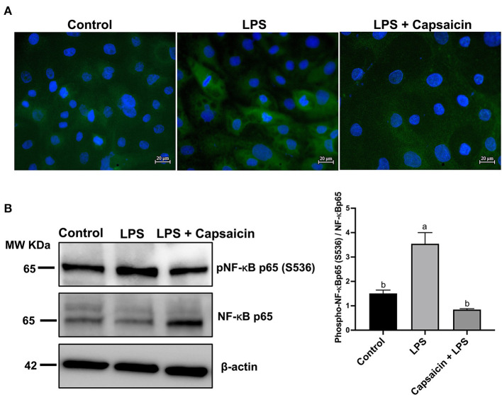
Figure 4.
Effects of capsaicin and LPS treatment on the phosphorylation of NF-κB p65 in LPS-challenged IPEC-J2 cells. The level of phosphorylated NF-κB p65 was evaluated using Immunofluorescence staining (A) and Western blotting (B), respectively. For Immunofluorescence staining, IPEC-J2 cells were seeded into coverslips at a density of 1×10⁵/well and cultured for 2 weeks. After being pretreated with capsaicin (100 μM), cells were stimulated with LPS. Cells then were then fixed for phosphorylated NF-κB p65 staining as described in the Materials and Methods. As for Western blot, IPEC-J2 cells were cultured in 6-well plates. Cells were firstly pre-treated with capsaicin (100 μM) for 2 h and then stimulated with LPS (10 μg/mL) for 6 h. The protein was extracted and the total NF-κB p65, as well as phosphorylation of NF-κB p65 in LPS-challenged IPEC-J2 cells were detected as described in the Materials and Methods. Values are presented as mean ± SEM. Different letters on bars (a, b) indicate significant differences, P < 0.05.

