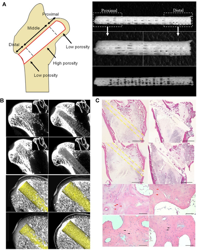
FIGURE 7.
FGS and FGS/BMMC could promote bone regeneration in the area of osteonecrosis. (A) A schematic image shows how the three segments of FGS with different porosities are distributed in the femoral head. The FGS consisting of three segments of spatially graded porosity, including 4 mm length proximal segment of 15% porosity, 17 mm length middle segment of 40% porosity, and 6 mm distal segment of 15% porosity. (B) FGS is degraded at the proximal end, and there is mineralized tissue around it. (C) Histological analysis confirmed that there was more new bone formation around FGS than other groups. Reproduced with permission (Maruyama et al., 2018). Copyright 2018, Elsevier Ltd.

