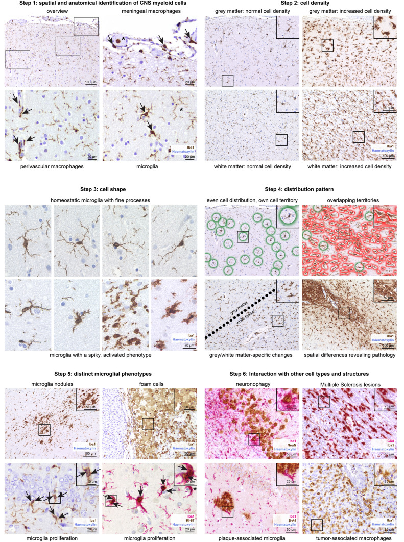
Fig. 3.
Immunohistochemistry-based assessment of microglia phenotypes across neuropathologies. Using Iba1 immunohistochemistry (brown), the following six-step standardized sequence is recommended for the analysis of microglia cells. In short, microglia cells and CNS-associated macrophages first need to be identified by their spatial distribution. Secondly, the cell density should be evaluated. The cell shape is of particular importance when analyzing the microglial phenotype. An uneven, irregular distribution pattern and microglia cell territories that are potentially overlapping might point towards local pathological processes within a tissue section. Next, the examiner should look for distinct microglia phenotypes, e.g. dividing cells or cells that underwent foam cell transformation. Lastly, a potential interaction with other cell types should be explored. Combined immunohistochemical reactions with two different markers are indicated. Counterstaining with haematoxylin (blue)

