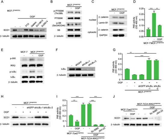
Figure 7.

OGP suppresses SCD1 expression by inhibiting the NF‐κB pathway. A) MCF‐7mammo cells preincubated with or without indicated inhibitors were treated with OGP. The level of SCD1 was detected by western blotting (n = 3). B,C) MCF‐7mammo cells were treated without or with OGP. B) Levels of total p70 S6 Kinase (P70S6K), S6 Ribosomal Protein (S6), and phosphorylated p70 S6 Kinase (p‐P70S6K), S6 Ribosomal Protein (p‐S6) were determined by western blotting (n = 3). C) Levels of intranuclear and cytosolic β‐catenin were determined by western blotting (n = 3). D,E) MCF‐7 cells and MCF‐7mammo cells were treated without or with OGP. Nuclear proteins were analyzed for NF‐κB DNA binding activity by D) ELISA and phosphorylated IKK, and IκBα were detected by E) western blot analysis (n = 3). F) IκBα levels in MCF‐7mammo cells transduced with IκBα shRNA (n = 3). G,H) MCF‐7mammo cells with or without IκBα knockdown were treated with or without OGP. G) Nuclear proteins were analyzed for NF‐κB DNA binding activity by ELISA (n = 3). H) SCD1 levels were determined by western blotting (n = 3). I,J) MCF‐7mammo cells transfected with (CA‐IKK2) or without (wt) plasmid expressing CA‐IKK2 were preincubated without or with indicated inhibitors before OGP treatment. I) Nuclear proteins were analyzed for NF‐κB DNA binding activity by ELISA (n = 3). J) SCD1 levels were determined by western blotting (n = 3). D,G,I) Mean ± SEM, **P < 0.01; ***P < 0.001 by one‐way ANOVA.
