
Figure 3.
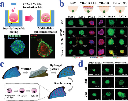
a) Schematic and fluorescence images of cell spheroid formation in NLM system created by superhydrophobic coating. Reproduced with permission.[ 140 ] Copyright 2019, American Chemical Society. b) Schematic diagram and representative fluorescence microscopy images of the different configurations for spheroids formed by hanging drop strategy. Reproduced with permission.[ 142 ] Copyright 2017, Wiley‐VCH. c,d) Schematic diagrams and fluorescent images illustrating the formation of cell spheroids on the superhydrophobic butterfly wing with hydrophilic hydrogel spots. The scale bar in (d) is 500 µm. Reproduced with permission.[ 143 ] Copyright 2019, American Chemical Society.