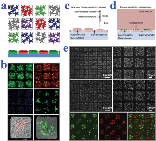
Figure 5.

a) Schematic representation of isolated transfected cell clusters in microspots without migration and cross‐contamination. b) Fluorescent images of four different cell lines cultured on the array and HEK cells transfected with different plasmids on two superhydrophilic regions. a,b) Reproduced with permission.[ 149 ] Copyright 2011, Wiley‐VCH. c,d) Schematic diagrams of the reverse transfection procedure using the superhydrophilic/superhydrophobic patterned surfaces. e) Microscopy images showing HEK 293 cells and HeLa cells confined in square superhydrophilic spots; fluorescent images of the transfected cells showing minimal cross‐contamination between neighboring regions. Reproduced with permission.[ 150 ] Copyright 2016, Wiley‐VCH.
