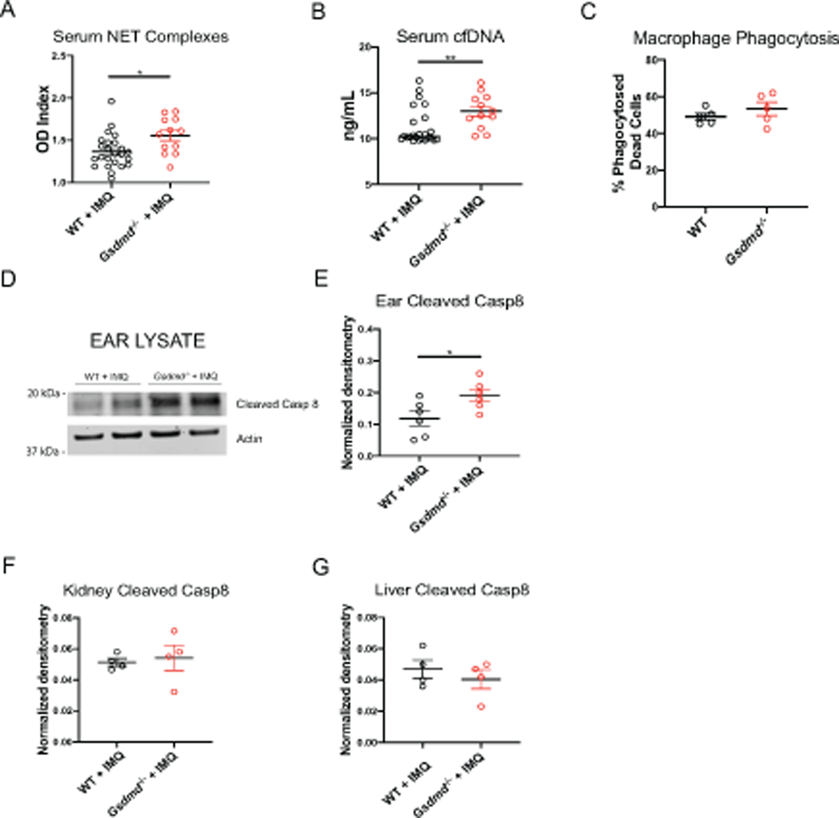
Figure 4. Gsdmd−/− mice display increased circulating autoantigens and enhanced release of HMGB1.

(A–B) Serum NET complexes and cell-free DNA (cfDNA) (n=24, WT + IMQ; n=12, Gsdmd−/− + IMQ). (C) Quantification of macrophage phagocytosis of apoptotic cells. (D) Immunoblot of cleaved caspase 8 in ear lysates of WT and Gsdmd−/− mice treated with imiquimod for 1 week. (E–G) Quantification of cleaved caspase 8 normalized to actin in ear, kidney, and liver lysates of WT and Gsdmd−/− mice treated with imiquimod for 1 week.
