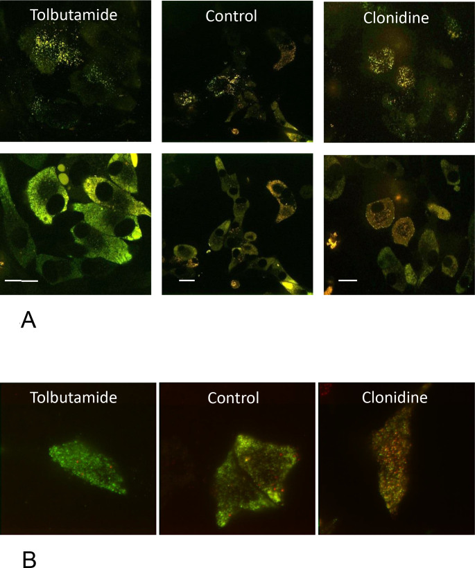
Figure 4.
Aspect of granulation state and size of MIN6 cells labeled with hIns-dsRed E5. (A) MIN6 cells labeled with hIns-dsRed E5 were cultured for 18 hours either in the presence of 500 µM tolbutamide (left) or 1 µM clonidine (right) or control cultured (middle) and imaged by SD-CLSM close to the plasma membrane (0.2 µm, upper row) or close to the cell middle (4 µm, lower row). Note the considerable mistargeting and heterogeneous appearance. The length of the bars is 10 µm. (B) In the TIRF mode, a clear identification of single granules was possible after culture in the presence of tolbutamide or clonidine or after control culture. The different ratio of green to red fluorescence is clearly visible. Note the presence of red granules in the submembrane space. SD-CLSM, spinning disk confocal laser scanning microscopy.

