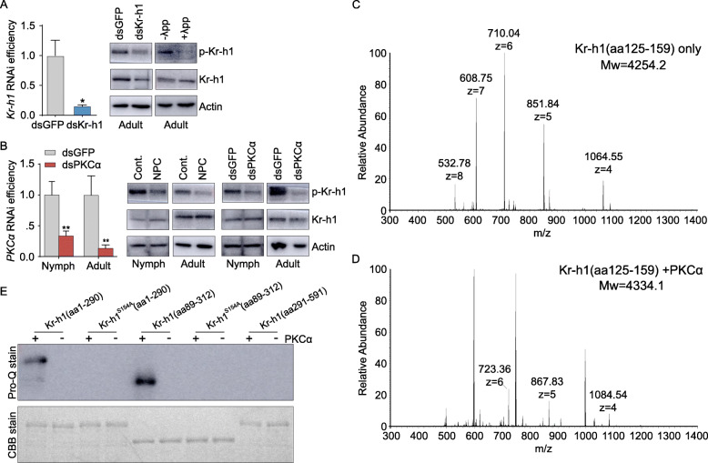
Fig. 1.
Phosphorylation of Kr-h1 by PKCα at Ser154. A Left panel: Kr-h1 RNAi efficiency in the fat body of 3-day-old adult females. *P<0.05. n=8. Right panel: Verification of phospho-Kr-h1 (Ser154) antibody specificity by using protein extracts from the fat body of 3-day-old adult females subjected to Kr-h1 knockdown and phosphatase λpp treatment. B Left panel: PKCα RNAi efficiency in the whole body of penultimate 4th instar nymphs and the fat body of 3-day-old adult females. **P<0.01. n=8. Right panel: Relative levels of Kr-h1 and phosphorylated Kr-h1 (p-Kr-h1) in the whole body of 4th instar nymphs and the fat body of 3-day-old adult females treated by NPC15437 (NPC) vs. DMSO solvent control (Cont.) and dsPKCα vs. dsGFP control. C-D LC-MS/MS analysis of wildtype Kr-h1(aa125-159) peptide (C) and Kr-h1(aa125-159) preincubated with PKCα (D). m/z indicates the mass to charge ratio. E Upper panel: Pro-Q Diamond Phosphoprotein Gel Stain of purified bacterially-expressed GST-tagged peptides of Kr-h1(aa1-290), Kr-h1S154A(aa1-290), Kr-h1(aa89-312), Kr-h1S154A(aa89-312), and Kr-h1(aa291-591) preincubated with or without PKCα. Lower panel: Coomassie brilliant blue staining was used as the loading controls

