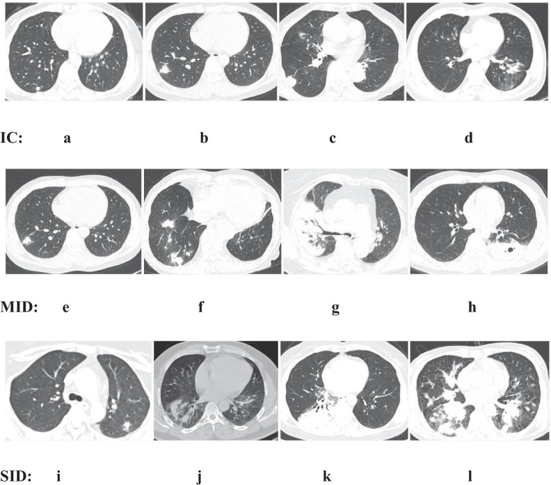
Fig. 5.
Typical chest computed tomography scans of cryptococcosis patients. IC: a-c: solitary subpleural nodules of varying sizes in the right lower lung (< 3 cm); d irregular nodular shadows in the left lower lung; MID: e solitary subpleural nodule with a cavity in the right upper lung; f multiple nodular shadows close to the periphery in the right lung; g consolidation of the right lung with air bronchogram sign; h lump in the lower left lung, with a cavity in the middle; SID: i solitary subpleural nodule in the left lung; j irregular nodular shadows in the right lower lung; k massive consolidation of the right lung with air bronchogram sign and a small amount of pleural effusion; l lump in the right lower lung with nodules on the other lobes

