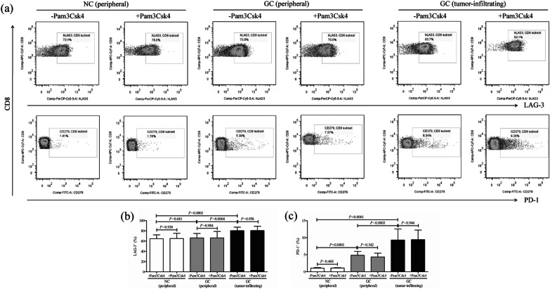
Fig. 4.
The influence of TLR2 activation on inhibitory molecules expression in peripheral and tumor-infiltrating CD8+ T cells from NC and GC patients. CD8+ T cells were purified from the same subjects as in Fig. 2, including peripheral bloods of NC (n = 9) and GC patients (n = 11), as well as from tumor residency of GC patients (n = 11), and were cultured for 12 h in the presence of anti-CD3/CD28 with or without TLR2 agonist Pam3Csk4. Lymphocyte activation gene-3 (LAG-3) and CD279 (programmed death-1, PD-1) expression in CD8+ T cells was measured by flow cytometry. a The representative flow dots of LAG-3 and PD-1 positive cells in peripheral CD8+ T cells from NC and GC patients, as well as in tumor-infiltrating CD8+ T cells from GC patents with or without Pam3Csk4 stimulation. The percentage of b LAG-3+ and c PD-1+ cells within CD8+ T cells was compared. The columns indicated means, and the bars indicated standard deviation. Significances were determined by LSD-t test or paired t test

