FIG. 7.
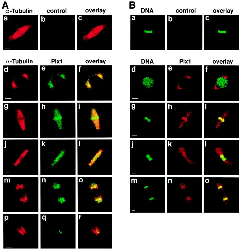
Localization of Plx1 changes during mitosis. (A) Late-blastula-stage embryos were fixed in methanol, and immunofluorescence staining was performed as previously described (30). α-Tubulin was detected with an anti-α-tubulin monoclonal antibody (Sigma) and visualized by Cy3-conjugated donkey anti-mouse IgG antibodies, and Plx1 was detected with anti-Plx1 antibodies and visualized by Cy2-conjugated donkey anti-rabbit IgG antibodies. Confocal microscopy was performed with an MRC-600 microscope (Bio-Rad). Bars, 10 μm. (B) Embryos were fixed as in panel A. Plx1 was visualized by Cy3-conjugated donkey anti-rabbit IgG antibodies and DNA was detected with SYTOX Green. Control IgG from immune sera that had been depleted of all Plx1-specific antibodies was used as a negative control (b).

