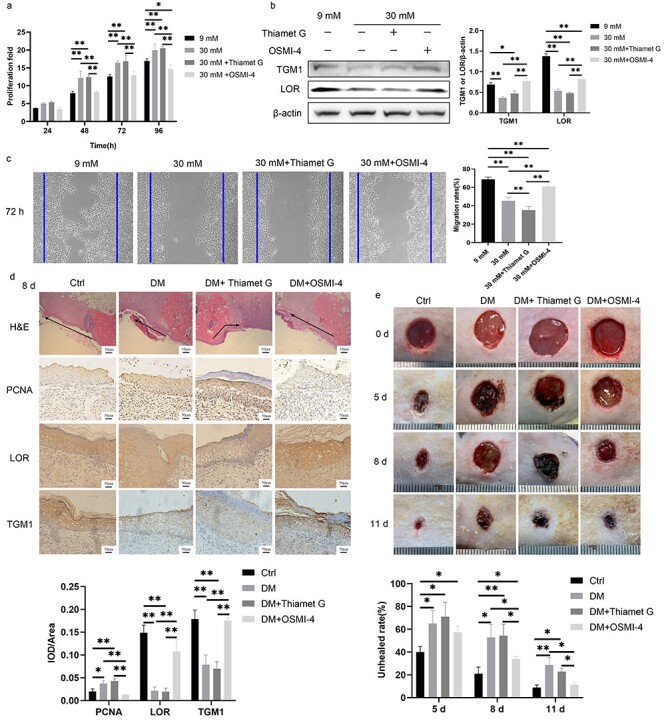
Figure 7.

Inhibition of O-GlcNAcylation promoted diabetic wound healing. (a) The proliferation of HaCaT cells treated with 9 mM glucose, 30 mM glucose, 30 mM + Thiamet G and 30 mM + OSMI-4 was demonstrated by CCK-8 analysis. (b) Expressions of TGM1 and LOR of HaCaT cells treated with 9 mM glucose, 30 mM glucose, 30 mM + Thiamet G and 30 mM + OSMI-4 were measured by Western blotting. (c) The migration of HaCaT cells treated with 9 mM glucose, 30 mM glucose, 30 mM + Thiamet G and 30 mM + OSMI-4 was detected by the scratch assay at 72 h. (d) Thiamet G (200 μL, 10 mg/mL) or OSMI-4 (200 μL, 10 mg/mL) was administered subcutaneously at the edge of the wound for 8 consecutive days in the DM + Thiamet G group or DM + OSMI-4 group, respectively. Migration, proliferation (PCNA), and differentiation (LOR and TGM1) of keratinocytes at rat wound margin of different groups were measured. The black arrow indicates the direction of epidermal migration. (e) Representative pictures of wound healing in rats from different groups and quantitative statistics of non-healing rate are shown. Scale bar: 10 μm, 50 μm. *p < 0.05, **p < 0.01. Ctrl control, DM diabetes mellitus, H&E hematoxylin and eosin staining, IOD integrated optical density, LOR loricrin, PCNA proliferating cell nuclear antigen, TGM1 transglutaminase 1
