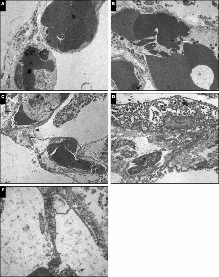
Figure 3.
Ultrastructural analyses (uranyl acetate/lead citrate). A, Patient 1. Alveolar capillaries are filled with erythrocytes and fragmentocytes (asterisks) embedded in an alveolar septum with slightly increased extracellular matrix accumulation (×4,000; image width, 23.5 µm). B, Patient 1. Capillaries are filled with erythrocytes and fragmentocytes (asterisk). The erythrocytes are altered in shape and form very long and thin cytoplasmic processes as a hint of a special kind of acanthocytosis (arrows) (×3,000; image width, 31.5 µm). C, Patient 2. Alveolar septum with erythrocytes and leukocyte-filled capillaries is enlarged by interstitial fluid accumulation and loosely organized fibrillar (arrows) and nonfibrillar extracellular matrix (arrowheads). The septum is bounded by alveolar basement membrane with sheared-off alveolar epithelium (×3,000; image width, 31.5 µm). D, Patient 3. Interstitial fibrosis of a thickened alveolar septum with fibroblast debris (asterisks) and an adjacent alveolar epithelial cell (a). The extracellular matrix contains collagen fibrils, elastin, and an accumulation of basement membrane material (×3,000; image width, 31.5 µm). E, Patient 3. The alveolar capillary endothelium reveals cytoplasmic processes with the formation of an interendothelial contact (arrows). The cytoplasmic process divides the lumen as a sign of intussusceptive (splitting) angiogenesis (×20,000; image width, 5.9 µm).

