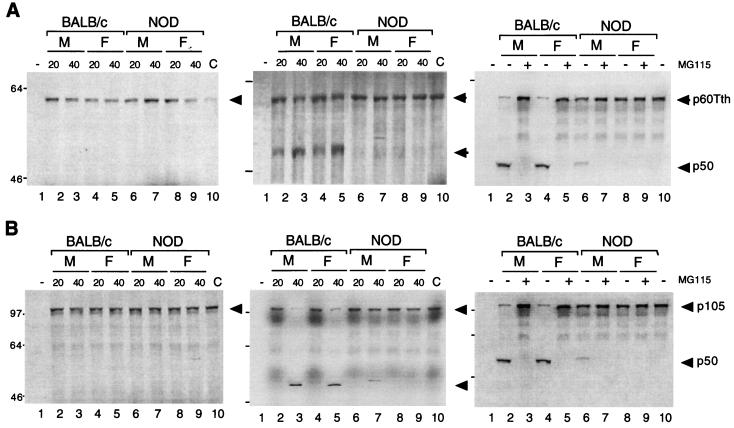
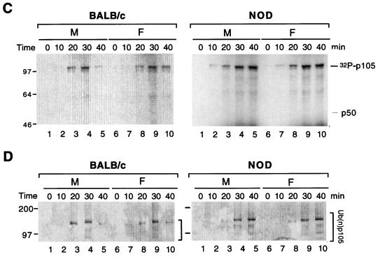
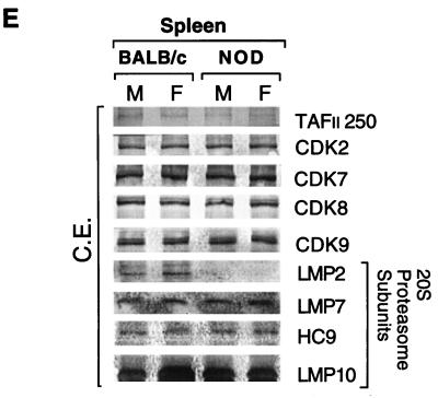
FIG. 4.
Proteolysis of p105 mediated by cytosolic extracts of BALB/c and NOD mice in vitro. (A and B) Purified 35S-labeled recombinant p60Tth (A) or p105 (B) was incubated in a reaction mixture containing cytosolic extract of male (M) or female (F) BALB/c or NOD mouse spleen cell (20 or 40 μg of protein in left and center panels and 40 μg of protein in right panel) in the absence (left panels) and presence (center and right panels) of 10 mM ATP. Incubations in the right panels were also performed in the absence (−) and presence (+) of 50 μM MG115. Lanes 1 correspond to reaction mixtures without substrate; lanes 10 correspond to negative controls without added extract. Incubations were at 30°C for 90 min, after which SDS-PAGE and autoradiography were performed. (C) Recombinant p105 was incubated for various periods of time at 30°C in a reaction mixture containing [γ-32P]ATP cytosolic extract (40 μg of protein) of spleen cells from male or female BALB/c or NOD mice, after which p105 was immunoprecipitated with antibodies to p50 and subjected to SDS-PAGE and autoradiography. The positions of phosphorylated p105 and unphosphorylated p50 are indicated. (D) Recombinant p105 was incubated for various time periods at 30°C in a reaction mixture containing cytosolic extract (40 μg of protein) of spleen cells from male or female BALB/c or NOD mice, after which complexes were cross-linked with glutaraldehyde, immunoprecipitated with antibodies to p50, and detected by immunoblot analysis with antibodies to ubiquitin. The positions of ubiquitinated p105 [Ub(n)-p105] and of molecular size standards (in kilodaltons) are indicated. (E) Immunoblot analysis of proteasome subunits in BALB/c and NOD mice. Cytosolic extract of spleen cells from male or female BALB/c or NOD mice was subjected to immunoblot analysis with 20S proteasome subunit antibodies and control CDK and TAFII250 antibodies.


