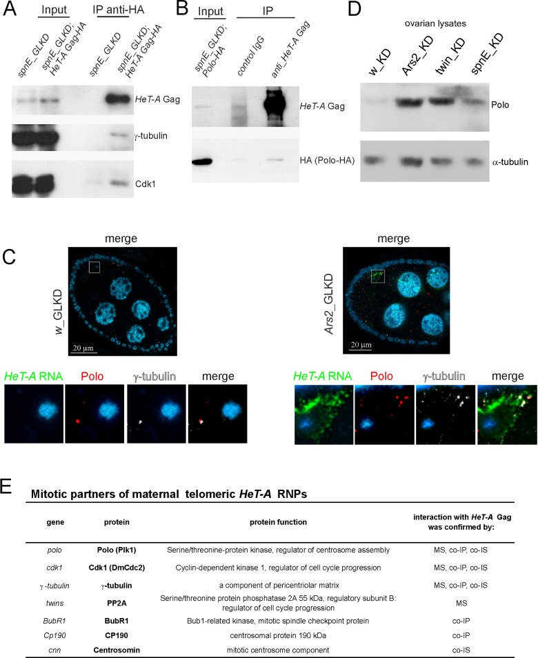
Fig 3. HeT-A ribonucleoprotein particles interact with Polo during oogenesis.
(A) A coimmunoprecipitation experiment was performed on ovarian extracts from spnE_GLKD flies expressing HeT-A Gag-HA. (B) A co-IP experiment was performed on ovarian extracts from spnE_GLKD flies expressing Polo-HA. The samples were separated by SDS-PAGE and analyzed by western blotting using the antibodies indicated on the right. The starting lysate (input) and immunoprecipitated (IP) probes are indicated; the antibodies used for co-IP are indicated above the IP lanes. (C) HeT-A RNA FISH (green) combined with immunostaining of Polo (phosphorylated form, red) and γ-tubulin (grey) in the control (left panel) and Ars2_GLKD ovaries (right panel). Egg chambers at 9–10 stages of oogenesis are shown. The lower panels show the enlarged areas highlighted by squares. The blue color indicates DNA. (D) Western blot analysis of ovary lysates probed with anti-Polo antibody; α-tubulin was used as a loading control. (E) Mitotic partners of maternal HeT-A RNPs revealed by different methods. MS, mass spectrometry; co-IP, co-immunoprecipitation; co-IS, co-immunostaining.

