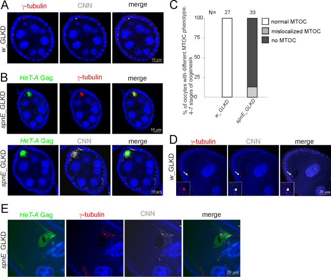
Fig 4. HeT-A overexpression caused by spnE knockdown is accompanied by centrosome dysfunction during oogenesis.
(A) Co-immunostaining demonstrates colocalization of CNN (grey) and γ-tubulin (red) in the control ovaries (w_GLKD). (B) Co-immunostaining demonstrates the localization of HeT-A Gag (green), CNN (grey), or γ-tubulin (red) in spnE_GLKD ovaries. Stage 6 of oogenesis is shown (A, B). (C) Bar diagrams show the MTOC phenotype (%) observed at 4–6 stages of oogenesis in spnE_GLKD. (D) Co-immunostaining of CNN (grey) and γ-tubulin (red) shows the centrosome reduction at stage 9 of oogenesis in w_GLKD ovaries. (E) Immunostaining reveals the accumulation of HeT-A Gag (green) and mislocalized CNN (grey) and γ-tubulin (red) at stage 10 of oogenesis in spnE_GLKD. The blue color indicates DNA.

