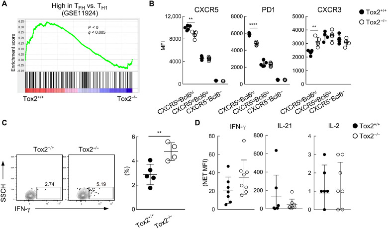
Fig. 6. GC TFH cells in Tox2-deficient mice exhibited less TFH gene signature.
(A) GSEA analysis in Tox2+/+ and Tox2−/− PD1hiCXCR5hi GC TFH cells with the gene set of up-regulated TFH compared to effector TH1. (B) MFI of CXCR5, PD1, and CXCR3 in CXCR5hiBcl6hi, CXCR5loBcl6lo, and CXCR5−Bcl6− CD4+ T cells. N = 4 to 5. (C) IFN-γ expression of spleen CD4+ T cells 7 days after H3N2 infection. Splenocytes were incubated with heat-inactivated H3N2 influenza virus for 12 hours followed by another 12-hour culture in the presence of GolgiPlug and brefeldin. IFN-γ expression by splenic CD4+ T cells was assessed by FACS. Representative flow data (left panel) and the dataset of four to five mice are shown (right). (D) IFN-γ, IL-21, and IL-2 amount in serum from mice 7 days after SRBC immunization. **P < 0.01; ****P < 0.001.

