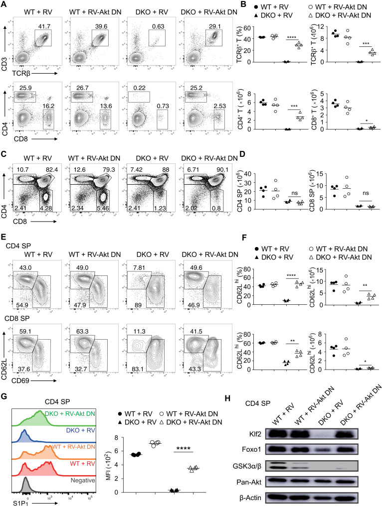
Fig. 5. Suppression of Akt signaling restores the egress of GSK3-deficient thymocytes.
(A) Flow cytometry analysis of CD3+TCRβ+ (top), CD4+, and CD8+ (bottom) T cells in the lymph nodes of recipient mice reconstituted with WT and DKO bone marrow cells transduced with empty retroviruses (RVs) or retroviruses encoding dominant negative Akt (RV-Akt DN). (B) Summary of the percentage and number of CD3+TCRβ+ (top), CD4+, and CD8+ T cells (bottom) in (A) (n ≥ 3 per group). (C) Flow cytometry analysis of thymocyte development in the recipients in (A). (D) Summary of the numbers of CD4 and CD8 SP thymocytes in (C). (E) Flow cytometry analysis of CD62LhiCD69lo, CD62LloCD69hi CD4 (top), and CD8 (bottom) SP thymocytes in the recipients in (A). (F) Summary of the percentage and number of CD62LhiCD69lo CD4 and CD8 SP thymocytes in (E). (G) S1P1 expression on CD62LhiCD69lo CD4 SP thymocytes in the recipients in (A) was analyzed by flow cytometry. CD62LloCD69hi cells as a negative control (gray). Right: MFI of S1P1 expression on CD62LhiCD69lo CD4 SP thymocytes of indicated groups. (H) Immunoblot analysis of Klf2, Foxo1, GSK3α/β, Akt, and β-actin in CD4 SP thymocytes from indicated groups. Each symbol represents an individual mouse. Small horizontal lines indicate the mean (± SEM). *P < 0.1; **P < 0.01; ***P < 0.001; ****P < 0.0001. Data are representative of two independent experiments.

