
Figure 4. Cellular risk factors associated with or proposed to influence HLA-restricted drug-induced T-cell activation.
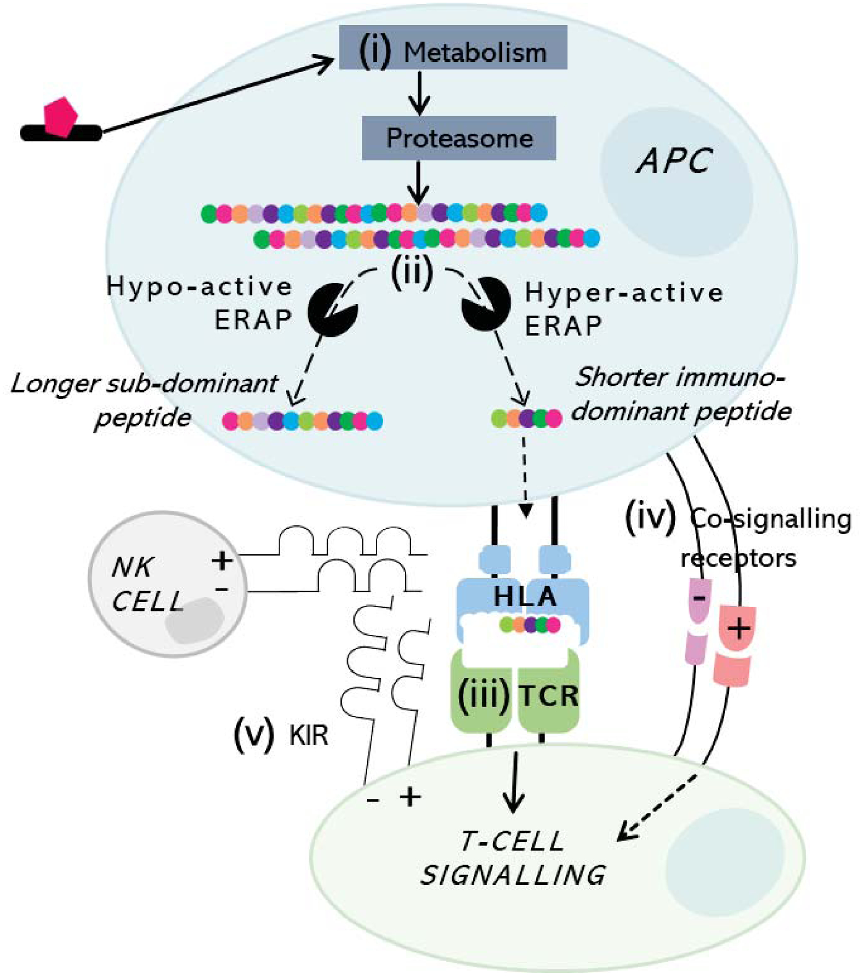
Antigen may undergo (i) metabolism and proteasomal processing to peptides which finally undergo trimming via (ii) endoplasmic reticulum aminopeptidases (ERAP) to an optimal length before loading into the HLA class I-binding groove. Risk HLA-presented peptide is then presented to T-cells with corresponding (iii) TCR specificity, forming signal 1 (stable HLA-antigen-TCR) in the presence of (iv) conversely co-signalling pathways skewing resultant response as signal 2 towards cytotoxicity (co-stimulation) or tolerance (co-inhibition). (v) KIR expressed on both NK- and T-cells bind to distinct HLA epitopes with specificity for presented peptide to regulate degranulation and cytotoxic response.