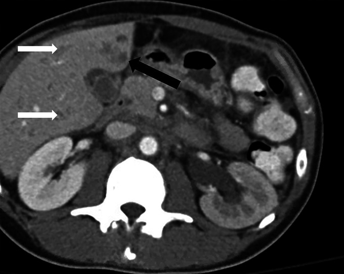
Figure 1.

Axial view of the abdominal and pelvic computed tomography with intravenous contrast demonstrating a 1.2 × 1.2-cm hypodense lesion (black arrow) in the anterior-inferior left hepatic lobe. Additional scattered subcentimeter hypodensities (white arrows) are also seen in the liver.
