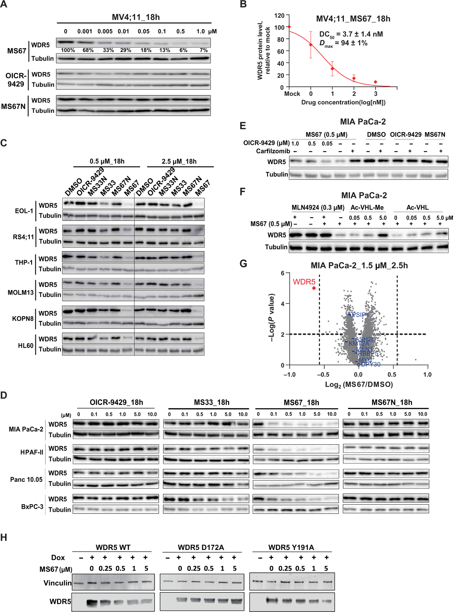
Fig. 4. MS67 potently and selectively degrades WDR5 in MLL-r AML and PDAC cells.

(A) Immunoblots for WDR5 and Tubulin posttreatment of MV4;11 cells with the indicated concentrations of MS67, MS67N, or OICR-9429 for 18 hours. (B) DC50 and Dmax values of MS67 in MV4;11 cells are shown as the means ± SD from three independent experiments. MV4;11 cells were treated with MS67 for 18 hours. The band intensity is determined by ImageJ software. (C) Immunoblots for WDR5 and Tubulin posttreatment of the indicated MLL-r AML cell lines and HL-60 (a non-MLL-r leukemia cell line) with dimethyl sulfoxide (DMSO) and 0.5 or 2.5 μM OICR-9429, MS33, MS33N, MS67, or MS67N for 18 hours. (D) Immunoblots for WDR5 and Tubulin posttreatment with the indicated concentrations of OICR-9429, MS33, MS67, or MS67N in the indicated PDAC cell lines for 18 hours. (E) Immunoblots for WDR5 and Tubulin after a 2-hour pretreatment with DMSO, carfilzomib (0.4 μM), or OICR-9429 (0.05, 0.5, and 1.0 μM), followed by a 4-hour treatment with 0.5 μM MS67 in MIA PaCa-2 cells. (F) Immunoblots for WDR5 and Tubulin after a 2-hour pretreatment with DMSO, MLN4924 (0.3 μM), or Ac-VHL-Me/Ac-VHL (0.05, 0.5, and 5 μM), followed by a 4-hour treatment with MS67 (0.5 μM) in MIA PaCa-2 cells. (G) Quantitative proteomics analysis of MIA PaCa-2 cells treated with 1.5 μM MS67 versus DMSO for 2.5 hours. A total of 4039 proteins were identified and quantified. The dashed lines indicate a cutoff of P value less than 0.01 (y axis) and fold change greater than 1.5 (x axis) in three biological replicates. (H) The effect of MS67 on degrading WDR5 WT and D172A and Y191A WDR5 mutants. HEK293T cells ectopically overexpressed with WDR5 WT and D172A and Y191A mutants, respectively, upon treatment with doxycycline (Dox) or DMSO, were treated with DMSO or MS67 at indicated concentrations for 72 hours.
