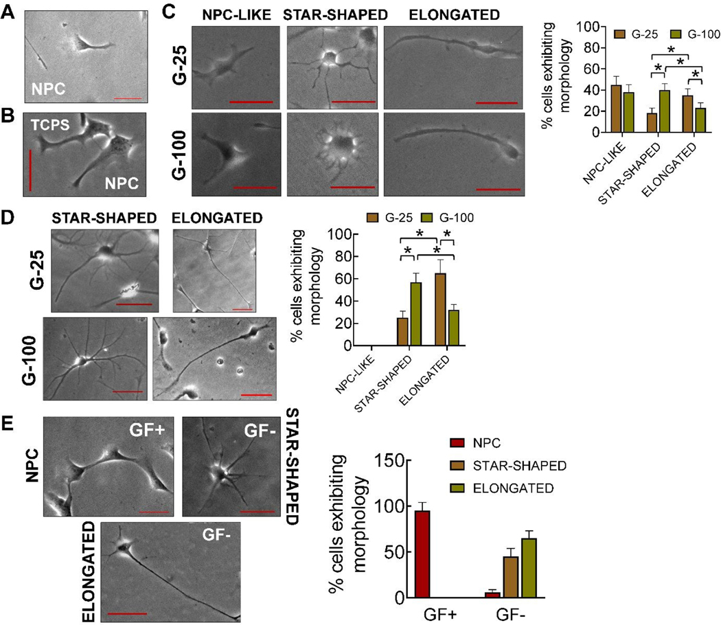
Fig 2.
Representative phase contrast images of (A) cells in complete medium (GF+), immediately after priming (three days in culture), (B) cells cultured on TCPS, three days post-priming, in the presence of complete medium, (C) NPC-like, star-shaped, and elongated cells on G-25 and G-100 after three days in culture post-priming, (D) star-shaped and elongated cells on G-25 and G-100 gels, nine days post-priming, in complete medium, and (E) cells after nine days of culture on laminin coated TCPS in the presence of complete medium or maintenance medium. Distinct morphologies of the cells in maintenance medium was evident unlike in the presence of complete medium. The number of cells exhibiting each morphology were counted manually from the images and normalized to the total cells counted in each case; n > 150 cells/case. * indicates p < 0.05 between respective cases. Scale bar: 50 μm.

