Figure 1.
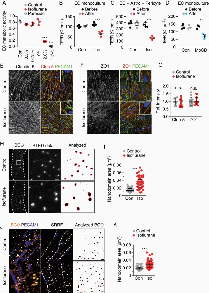
Isoflurane increases EC barrier permeability in vitro. (A) Mean viability ± SD of ECs treated with 0.5%–2% isoflurane (30 min) or peroxide as positive control (n = 3 experiments, 1-way ANOVA with Dunnett’s test), quantified by a WST1 assay that quantifies enzymatic activity of mitochondrial dehydrogenases present in viable cells. (B) Mean TEER ± SEM of EC cultures before and after 30 min 1% isoflurane treatment (n = 4 cultures, unpaired Student’s t-test). (C) Mean TEER ± SEM of triple cultures before and after 30 min 1% isoflurane treatment (n = 4 cultures, unpaired Student’s t-test). (D) Mean TEER ± SEM of EC cultures before and after 30 min 10 mM MbCD treatment (n = 3 cultures, unpaired Student’s t-test). (E–G) Co-immunofluorescence of PECAM1 with (E) claudin-5 and (F) ZO1 in EC cultures treated as in (B) (scale 20 µm) with (G) quantification of mean fluorescence intensity in n = 3 cultures. (H) STED images of ECs live-stained for cholesterol (BCtheta) after 30 min isoflurane treatment (cell borders, dashed line) with detailed area (scale 500 nm). Nanodomains included in the quantification are outlined in red (right). (I) Median size of BCtheta-positive nanodomains ± interquartile range (n = 50–51 cells from 3 experiments, Mann–Whitney test). (J) Confocal (left), chromatic aberration corrected SRRF (middle, cell borders, dashed line), and thresholded SRRF images (right) of endothelial cells treated as in (B) (scale 500 nm). (K) Median size of BCtheta-positive nanodomains ± interquartile range (n = 38–57 cells from 2 experiments, Mann–Whitney test).

