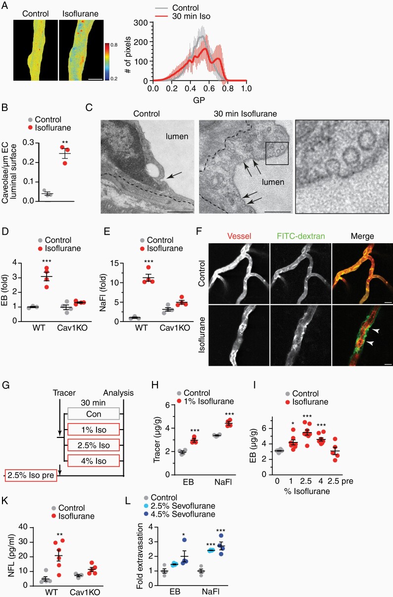
Figure 2.
Isoflurane disturbs BBB tightness in vivo. (A) Membrane fluidity of capillaries in mice treated with isoflurane (2.5%, 30 min) visualized by GP of Laurdan shown in false color with mean GP ± SEM of n = 13 vessels per condition. (B) Mean density of luminal micro-invaginations ± SEM in mice exposed to isoflurane (39 and 41 capillaries from n = 3 animals, unpaired Student’s t-test). (C) Electron micrographs of caveolar profiles (arrows) in brain ECs (dashed line, abluminal EC membrane), given in detail in boxed area (scales 500 nm). (D) Mean fold extravasation of EB ± SEM in WT and caveolin-1 deficient (Cav1KO) mice treated with isoflurane (n = 3–4 animals, 2-way ANOVA with Tukey’s test). (E) Mean fold extravasation of NaFl ± SEM in WT and Cav1KO mice treated with isoflurane (n = 3–4 animals, 2-way ANOVA with Sidak’s test). (F) Representative images of FITC-dextran extravasation in mice exposed to isoflurane (arrowheads). DiI-mediated vessel paint stained capillaries (scale 25 µm). (G) Experimental paradigm of BBB permeability. Mice were exposed to increasing dosages of isoflurane directly after tracer injection. In the 2.5% Iso pre-paradigm, tracer was administered to mice following the isoflurane treatment. (H) Mean extravasation of tracers (µg/g brain weight) ± SEM (n = 4 animals, 1-way ANOVA with Dunnett’s test). (I) Mean extravasation of EB (µg/g brain weight) ± SEM (n = 5–10 animals, 1-way ANOVA with Dunnett’s test, indicated are significant changes to controls). (K) Mean serum NFL ± SEM in WT and Cav1KO mice treated with 2.5% isoflurane for 30 min (n = 3–6 animals, 2-way ANOVA with Sidak’s test).

