. 2021 Sep 14;10:e72419. doi: 10.7554/eLife.72419

Figure 5. Myogenic fusion proteins are detected along filopodia in differentiating muscle cells.

(A) Immunofluorescent detection of Myomaker and Myomixer in differentiating myoblast cultures. Inset shows Myomixer and Myomaker puncta on the surface of a lamellipodial extension with filopodia. (B) Myomaker and Myomixer puncta are found localized to class X myosin (Myo10)-filled filopodia of differentiating myoblasts (days 4–5). Co-localization of (C) Myomixer and (D) Myomaker with Myo10 in filopodia remnants of the differentiating myoblast insoluble fraction (indicated with arrows). (E) Tibialis anterior (TA) muscle cross-sections of Pax7-WT mice at 4 days following cardiotoxin (CTX)-induced injury. (F) Cross-section (left) and longitudinal section (right) of Pax7-M10cKO TA muscle at 4 days following CTX-induced injury. Scale bars represent 10 µm.

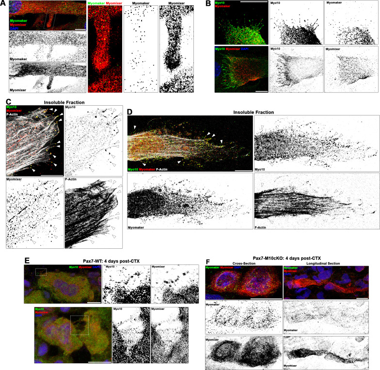
Figure 5.

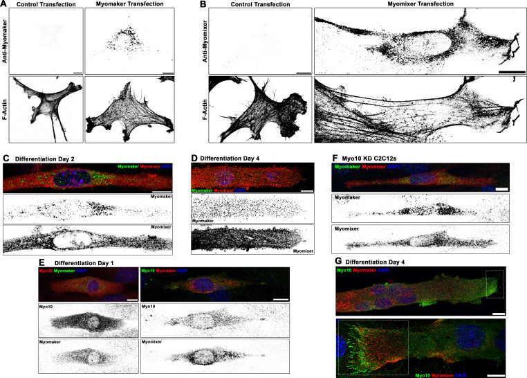
Figure 5—figure supplement 1. Localization patterns of Myomaker and Myomixer in differentiating myoblasts.

Figure 5—figure supplement 1.

Specificity testing of commercially available (A) Myomaker and (B) Myomixer antibodies for immunofluorescent detection of these proteins following transfection of undifferentiated myoblasts. Immunofluorescence (IF) staining pattern of Myomaker and Myomixer (C) early in myogenic differentiation and (D) in fully differentiated myotubes. Myomixer antibody was applied prior to cell permeabilization. (E) At day 1 of differentiation, myoblasts co-expressing class X myosin (Myo10) with Myomaker and Myomixer are observed. (F) Differentiated Myo10 knockdown (KD) cells express both Myomaker and Myomixer. (G) Localization patterns of Myo10 with Myomaker (top) and Myomixer (bottom) in differentiated myotubes (insets are found in Figure 5B). Scale bars represent 10 µm.
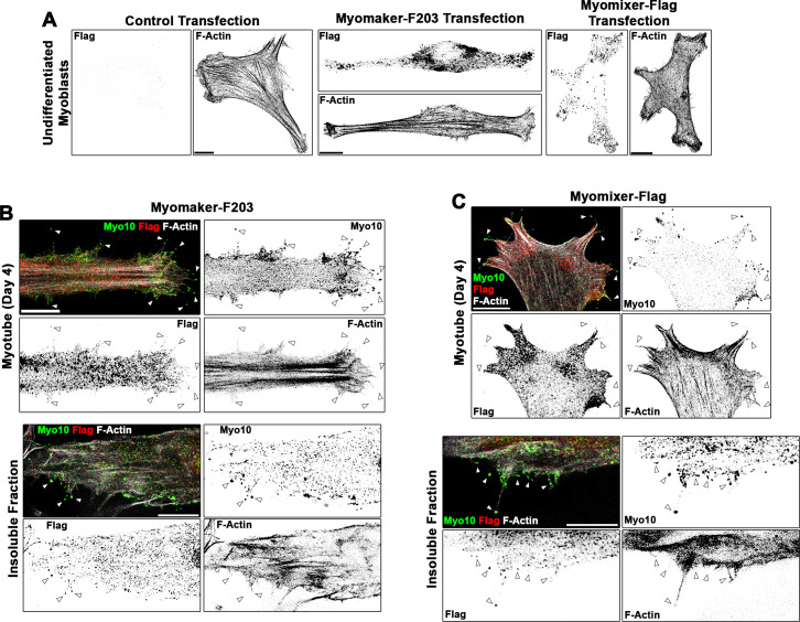
Figure 5—figure supplement 2. Localization patterns of Flag-tagged Myomaker and Myomixer constructs in differentiating myoblasts.

Figure 5—figure supplement 2.

(A) Anti-Flag staining of undifferentiated myoblasts following transfection of control, Myomaker-F203, or Myomixer-Flag plasmids reveals specificity of the anti-Flag antibody and that these Flag-tagged versions of Myomaker and Myomixer localize similarly to their wild-type counterparts (shown in Figure 5—figure supplement 1). Investigation of the localization of (B) Myomaker-F203 and (C) Myomixer-Flag in differentiated myotubes reveals Flag-positive puncta observed in class X myosin (Myo10)-positive filopodia of both whole cells and the insoluble cellular fraction. Scale bars represent 10 µm.