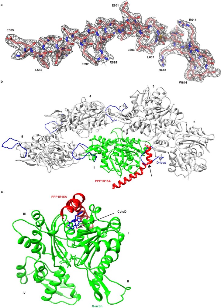
Extended Data Fig. 1. PPP1R15A binding to G-actin is exclusive to F-actin formation and CytoD binding.
(a) Stick diagram of PPP1R15A and the corresponding 2Fo-Fc map, contoured at 1.0σ within 2.0 Å of PPP1R15A atoms. (b) Ribbon diagram of F-actin (PDB 3J8A, tropomyosin omitted) in grey with protomers numbered. The PPP1R15A/G-actin complex (in red and green respectively, with DNase I removed) has been aligned with protomer 1. Actin D-loops are shown in blue and the clash between D-loop insertion and PPP1R15A binding to G-actin is indicated by the arrow. (c) The PPP1R15A/G-actin complex with actin domains numbered and a docked molecule of cytochalasin D (from PDB 3EKS), clashing with the C-terminal helix of PPP1R15A.

