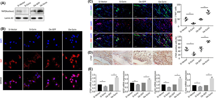
FIGURE 4.

(A) The Western blot was used to measure YAP protein expression in HUVECs n nucleus and cytoplasm following transfection and silencing of Ezrin. (B) Representative images of nuclear translocation of YAP. (C) Representative immunofluorescence staining images and positive cells of CD31 and YAP in synovial tissues of AIA‐treated mice after intraarticular injection of adenovirus. (D) CTGF protein levels in the IHC analysis and positive cells of the knee joints from inhibition and overexpression of Ezrin AIA mice (Scale bar, 25 μm). (E) The qPCR analysis of CTGF, bFGF, Cyr61 and C‐myc mRNA expression. n = 5, *p < 0.05, **p < 0.01
