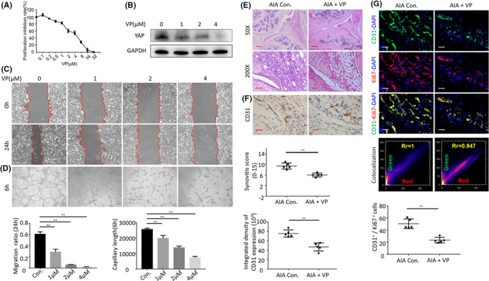
FIGURE 5.

(A) Inhibition of proliferation of verteporfin was measured with MTT assay. (B) The Western blot results showed that the verteporfin treatment inhibited the protein expression of YAP in HUVECs. (C) Representative images of wound‐healing assay. (D) Tube formation assay of HUVECs treated with verteporfin. (E) Representative images of haematoxylin and eosin staining in verteporfin treatment on AIA mice (Scale bar, 50 μm or 100 μm). (F) CD31 detection in the IHC analysis and positive area of the knee joints from treated AIA mice (Scale bar, 25 μm). (G) Representative immunofluorescence staining images and CD31&Ki67 colocalization, positive cells of CD31 and Ki67 in synovial tissues of treated AIA mice (scale bars, 50 μm and 25 μm). n = 5, *p < 0.05, **p < 0.01
