
FIGURE 5.
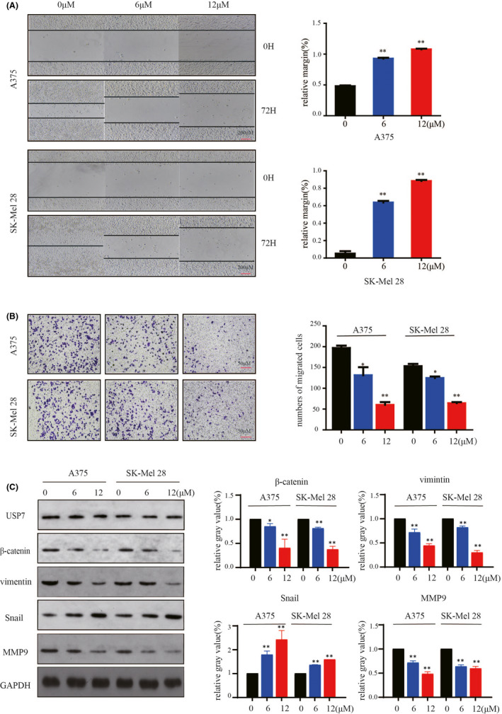
P22077 inhibits metastasis and invasion in melanoma cells. (A) A375 and SK‐Mel‐28 cells were treated at various concentrations of P22077 or DMSO. Then, the plates were scratch‐wounded with a 200‐μl sterile pipette tip and then incubated. The healing was recorded with a phase‐contrast microscope after 72 h. Right panel: quantitative analysis of relative margin(%) (mean values ± SEM, n = 3). Significant differences were evaluated using a one‐way ANOVA. *p < 0.05 vs control. (B) Transwell invasion assay determination of the number of melanoma cells that crossed the Matrigel layer after being treated with P22077 or DMSO (control). Right panel: the results represent the mean (n = 3) ± SD of each group, and significant differences were evaluated using Student's t test. *p < 0.05 vs 0 μM. (C) A375 and Sk‐Mel‐28 cells were treated with P22077 for 48 h, and Western blotting analysis of the expression of USP7 and some metastasis markers (β‐catenin, vimentin, snail, MMP9). Right panel: the results represent the mean (n = 3) ± SD of each group, and significant differences were evaluated using Student's t test. *p < 0.05 vs 0 μM