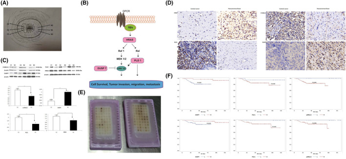
FIGURE 1.

Proteomic profiles of CC and paracancerous tissue and the identification of DEPs. Sample was collected according to the ‘sandwich’ method and obtained from 3 consecutive sites of suspicious lesions (C1/C2/C3) and normal‐looking areas (N1/N2/N3). If the specimens at both ends (C2/C3, N2/N3) were consistently confirmed by pathological examination as cervical invasive carcinoma and normal cervical tissue, respectively, the middle specimens (C1 and N1) were qualified (Figure 1A). The interaction of HRAS, DUSP7, PLD1 and p‐ERK1/2 is shown in Figure 1B. WB (Figure 1C) and IHC (Figure 1D) staining analyses consistently confirmed the results of the quantitative proteomic analysis that DEPs (HRAS, P‐ERK1/2 and PLD1) levels were increased, whereas the DUSP7 level was decreased in CC tissue compared with the paired normal paracancerous tissue. A total of 102 patients’ FFPE samples were included in the TMA (Figure 1E; optical magnification*20). The IHC results from the CC TMA analysis showed that the decreased expression of DUSP7 and increased expression of PLD1 and p‐ERK1/2 were adversely related to patients’ relapse (p = 0.003, 0.040 and 0.001, respectively; Figure 1F) and survival (p = 0.034, 0.001 and 0.006, respectively). *p < 0.05, **p < 0.01
