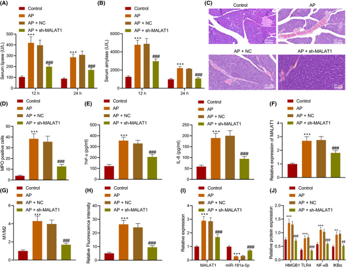
FIGURE 6.

Silencing of MALAT1 reduced pancreatic tissue injury in AP mice. AP mice were injected with sh‐MALAT1 (n = 8). A, Determination of serum amylase and lipase in AP mice. B, Determination of lipase in AP mice. C, Pancreatic tissue morphology in AP mice detected by H&E staining, scale bar = 25 μm. D, MPO expression in pancreatic tissues of AP mice. E, Levels of IL‐6 and TNF‐α in pancreatic tissues of AP mice measured by ELISA. F, MALAT1 expression in the serum‐derived EVs determined using RT‐qPCR. G, The phenotype of peritoneal macrophages detected by flow cytometry. H, The M1/M2 ratio detected by flow cytometry. H, M1 macrophages in pancreatic tissues detected by immunofluorescence staining. I, Expression of MALAT1 and miR‐181a‐5p in the pancreas of AP mice determined by RT‐qPCR. J, Protein levels of HMGB1, TLR4, NF‐κB and IKBa in the pancreas of AP mice determined by Western blot analysis. * vs. normal mice; # vs. AP mice or AP mice injected with NC. * or # p < 0.05. ** or ## p < 0.01. *** or ### p < 0.001. Data are shown as the mean ± standard errors
