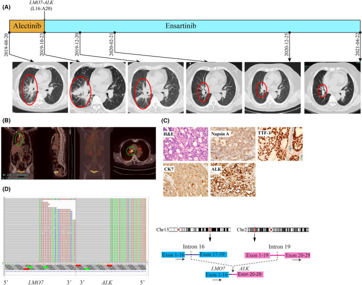
FIGURE 2.

Schematic of treatment history and NGS‐detected ALK fusion of Case 2. (A) The timeline of treatment and CT scans are showed where the red circles highlight the location of tumours. The time point of NGS‐detected LMO7‐ALK is labelled above. (B) Baseline positron emission tomography (PET‐CT) scans with circled tumour location. (C) Pathological examination of the surgical specimen. The H&E staining image (400×) showed a poorly differentiated adenocarcinoma histology, and IHC testing results showed positive expression of Napsin A, TTF‐1, CK7 and ALK respectively. (D) Identification of the LMO7‐ALK fusion. Sequencing reads of ALK and LMO7 are visualized by the Integrative Genomics Viewers (IGV, left). The schematic on the right shows the fused exons of the LMO7‐ALK rearrangement
