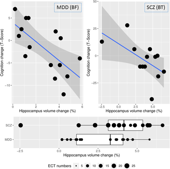
Fig. 3. The relationship between hippocampus volume change and cognitive performance change.
Left upper panel: Cohort with MDD patients and BF electrode placement, r = −0.68, df = 13, p = 0.005. Right upper panel: Cohort with schizophrenia patients and BT electrode placements, r = −0.58, df = 11, p = 0.04, (one individual could not participate in baseline cognitive testing). Lower panel: Patients with depression had on average 8.0 ± 0.6 ECTs between image acquisitions showed an average of 2.68% increase in hippocampal volume, patients with schizophrenia, who had on average 17.3 ± 3.4 ECTs between image acquisitions showed an average of 4.43% increase in hippocampus volume.

