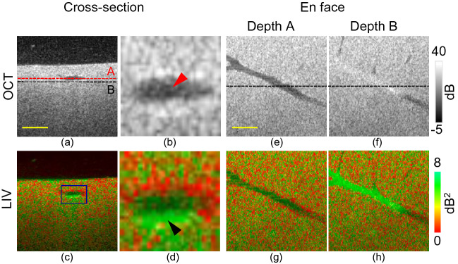
Figure 1.
Dynamics imaging of healthy mouse liver at the initial (0 h) time point. (a, c) Cross-sections from scattering OCT and LIV imaging; (b, d) magnified images of the cross-sectional images at the region indicated by the rectangular box in (c); (e, g) en face slices of OCT and LIV images at the depth location indicated by the red horizontal line in (a); (f, h) en face slices at the depth location indicated by the black horizontal line in (a). Scale bar: 250 μm.

