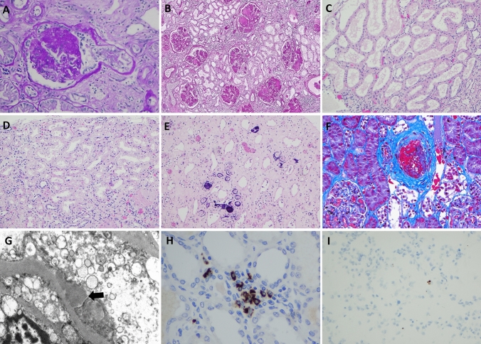
Fig. 1.
Histologic findings in kidney from postmortem patients with COVID-19. A Segmental obliteration of capillary tufts and prominence of epithelial cells consistent with focal segmental glomerulosclerosis (FSGS) (PAS × 200). B Diffuse mesangial nodular sclerosis in a case of diabetic nephropathy (PAS × 100). C Proximal tubules show diffuse attenuation of epithelial cells consistent with acute tubular injury (H&E × 100). D Acute tubular injury associated with vacuolization of proximal tubules (H&E × 200). E Calcium phosphate crystals identified within tubules (H&E × 100). F An arcuate artery shows bright red intraluminal thrombus (Gomori Trichrome × 200). G Electron microscopy shows subepithelial immune-type electron dense deposits (Transmission electron microscopy × 7000). H IHC stain for SARS-COV-2 spike protein showing granular staining of mononuclear cells in peritubular capillaries (× 200). I Rare positive staining of SARS-CoV-2 by RNAscope (× 100)

