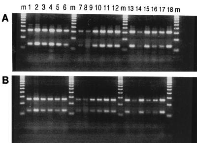
FIG. 1.
PRA gel after BstEII (A) and HaeIII (B) digestion of a 439-bp DNA fragment amplified from M. leprae bacilli isolated from various samples. Lanes m, molecular weight marker; lanes 1 to 5, samples from armadillos AV, AP, AJ, AU, and A10, respectively, calibrated to a 1:100 dilution; lanes 6 to 8, samples from nude mouse L1 calibrated to 1:100, 1:1,000, and 1:10,000 dilutions, respectively; lanes 9 and 10, a sample from nude mouse L1 that was purified to remove all host tissues and calibrated to a 1:100 dilution compared to the dilution of the same sample run in parallel as a partially purified preparation containing a significant amount of host tissues; lanes 11 to 13, samples from armadillos AU, AV, and AP, respectively, that were calibrated to 1:100 dilutions and that were gamma irradiated with 106 rads; lanes 14 and 15, a gamma-irradiated preparation from nude mouse L2 at 1:1,000 and 1:100 dilutions, respectively; lanes 16 to 18, gamma-irradiated preparations from nude mice L4, L3, and L1, respectively, at a 1:100 dilutions. See the text and Table 1 for details.

