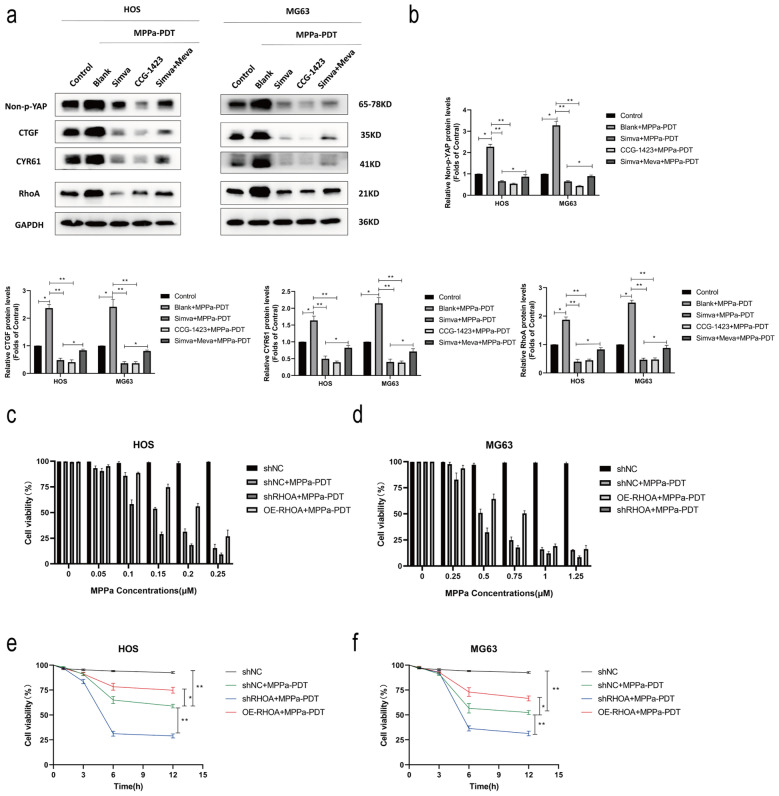
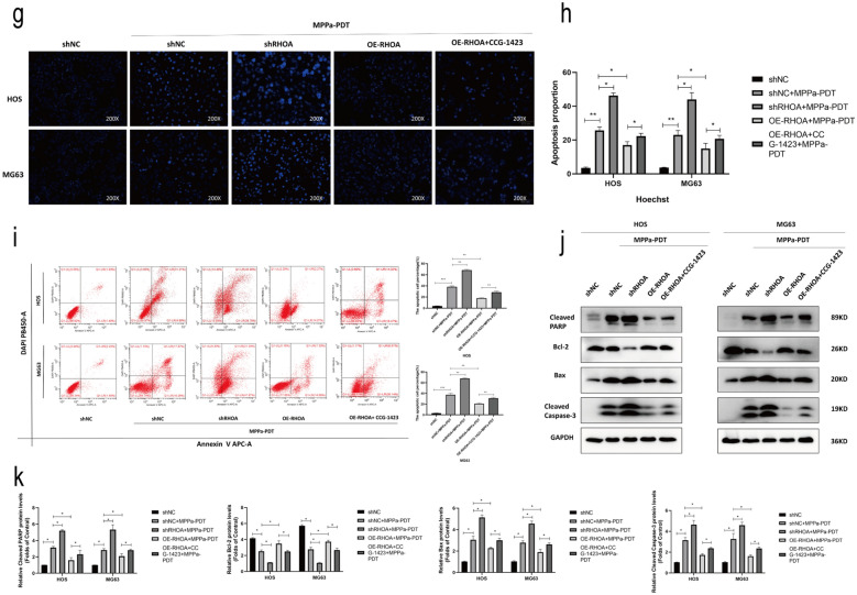
Fig. 5.
RhoA promotes OS cell resistance to MPPa-PDT. a, b Prior to MPPa-PDT, HOS and MG63 cells were treated with mevalonate (200 mM) for 6 h, simvastatin (4 mM for HOS, 8 mM for MG63) for 24 h, or CCG-1423 (5 μm) for 48 h, respectively. Western blotting was used to assess protein levels at 12 h post-MPPa-PDT, with GAPDH as a loading control. c, d Six different human OS cell lines (HOS-shNC, HOS-shRHOA, HOS-OE-RHOA, MG63-shNC, MG63-shRHOA, and MG63-OE-RHOA) were treated with various concentrations of MPPa for 20 h and then subjected to PDT, with a CCK-8 assay being used to assess cell viability at 12 h post-MPPa-PDT treatment. e, f Changes in OS cell viability following RhoA knockdown or overexpression were assessed at 12 h post-MPPa-PDT. g, h Hoechst staining demonstrating changes in OS cell apoptosis following RhoA knockdown or overexpression at 12 h post-MPPa-PDT. CCG-1423 treatment (5 μm for 48 h) was able to rescue the inhibition of apoptosis caused by the overexpression of RhoA. i HOS and MG63 cell apoptosis rates were assessed via flow cytometry. j, k Western blotting analyses of protein levels at 12 h post-MPPa-PDT treatment, with GAPDH as a loading control. Experiments were repeated in triplicate. * P < 0.05 **P < 0.01 ***P < 0.001


